What Is a Duplex Ultrasound?
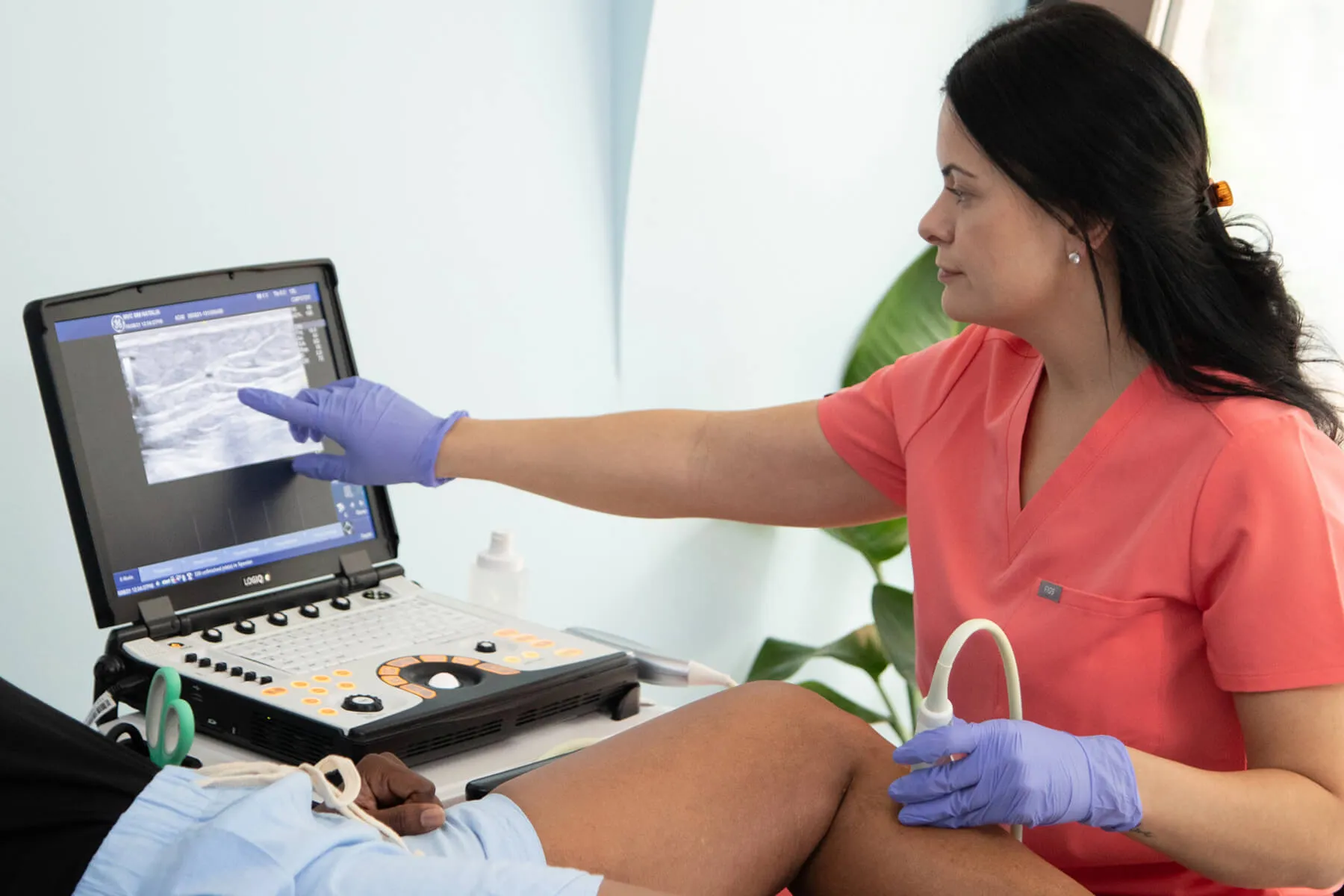
A duplex ultrasound is a painless, non-invasive scan that combines standard ultrasound imaging with Doppler to show both the structure of leg veins and the direction and speed of the blood flow. The term “duplex” means it uses two technologies at once: a 2-D image of the body’s internal structures and a Doppler ultrasound.
Unlike a general physical exam or other diagnostic procedures, a duplex ultrasound provides a direct look underneath the skin, making it a first-line test that vein specialists use to diagnose vein disease.

Types of Ultrasound Used to Evaluate Leg Veins
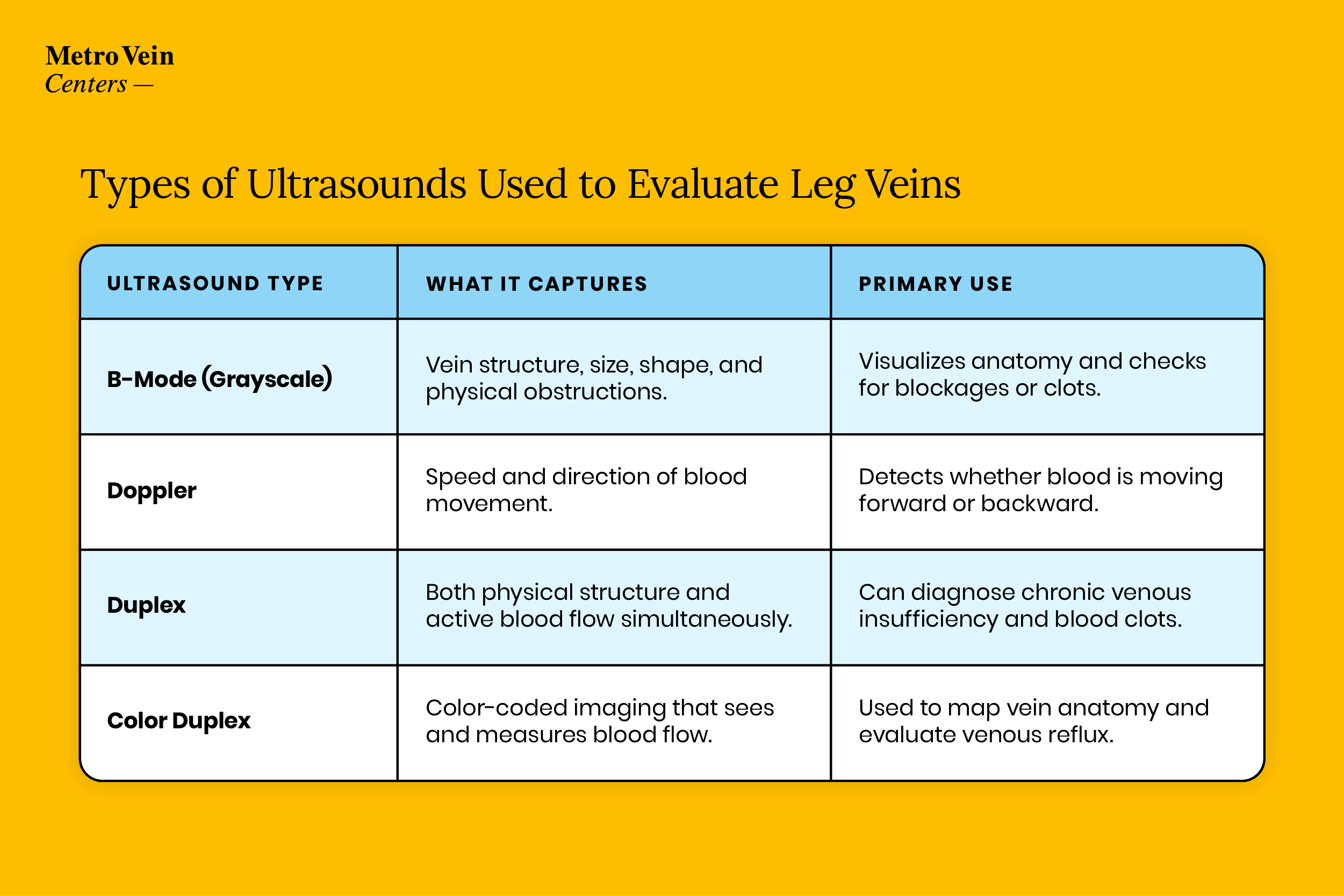
When looking into your diagnostic options, you may hear about three types of scans: B-mode, Doppler, and duplex ultrasound. Of these, duplex ultrasound is the primary scan used for vein disease because it provides a complete picture of both vein anatomy and blood flow.
Remember that single-mode scans can answer only one question: “what does the vein look like?” or “how is blood moving?” Only a duplex scan can answer both.
| Ultrasound Type | What It Captures | Primary Use |
|---|---|---|
| B-Mode (Grayscale) | Vein structure, size, shape, and physical obstructions. | Visualizes anatomy and checks for blockages or clots. |
| Doppler | Speed and direction of blood movement. | Detects whether blood is moving forward or backward. |
| Duplex | Both physical structure and active blood flow simultaneously. | Can diagnose chronic venous insufficiency and blood clots. |
| Color Duplex | Color-coded imaging that sees and measures blood flow. | Used to map vein anatomy and evaluate venous reflux. |
What Does a Duplex Ultrasound Show?
A duplex ultrasound can reveal important details about your vascular health, helping your doctor look beneath the surface to evaluate:
- Vein compressibility: Testing whether the vein walls collapse easily under light pressure (failure to collapse is a key indicator used to rule out or identify a blood clot).
- Valve function (reflux): Checking whether your vein valves close tightly or allow blood to leak backward due to gravity (identifying chronic venous insufficiency).
- Vein mapping: Pinpointing the exact layout and dimensions of your veins to plan targeted treatments like sclerotherapy or endovenous laser ablation (EVLA).
- Superficial, deep, and perforator system assessment: Examining all three layers of the leg’s venous network to find the source of the problem.
Reflux Time – How Results Are Measured
During an ultrasound, the technician tests for reflux by applying gentle pressure or asking you to flex your muscles, measuring how long gravity pulls blood backward before the valves catch. This duration is called reflux time.
A normal reflux time is under 0.5 seconds for superficial veins and under 1.0 second for deep veins.
Who Needs a Duplex Ultrasound?
Doctors typically order a venous duplex ultrasound for a patient when:
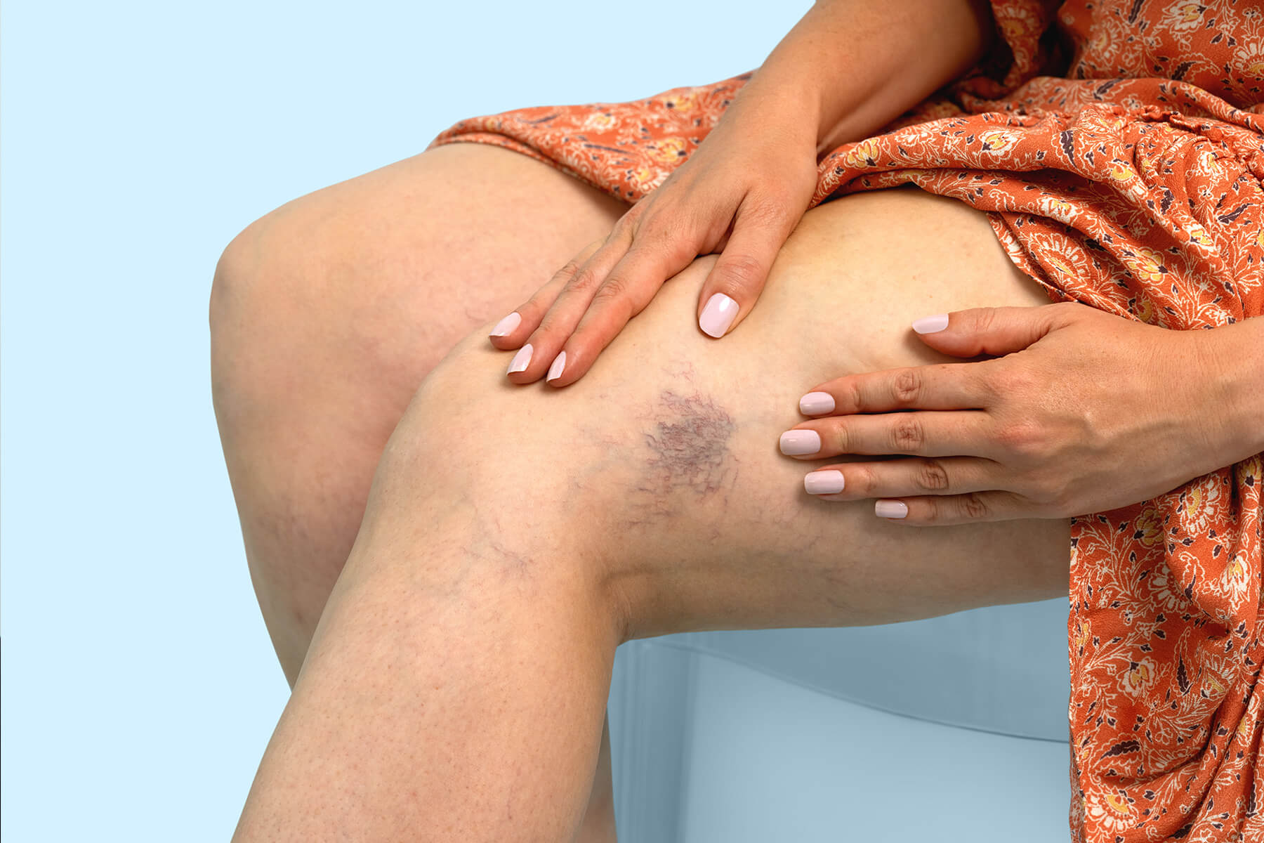
- They are assessing visible varicose veins or spider veins before starting treatment.
- A patient is experiencing uncomfortable leg symptoms like persistent swelling, aching and heaviness, or skin discoloration.
- They are checking for deep vein thrombosis (DVT)—marked by sudden swelling, warmth, or localized pain.
- They are performing post-treatment follow-ups after a prior vein procedure.
What to Expect During the Exam
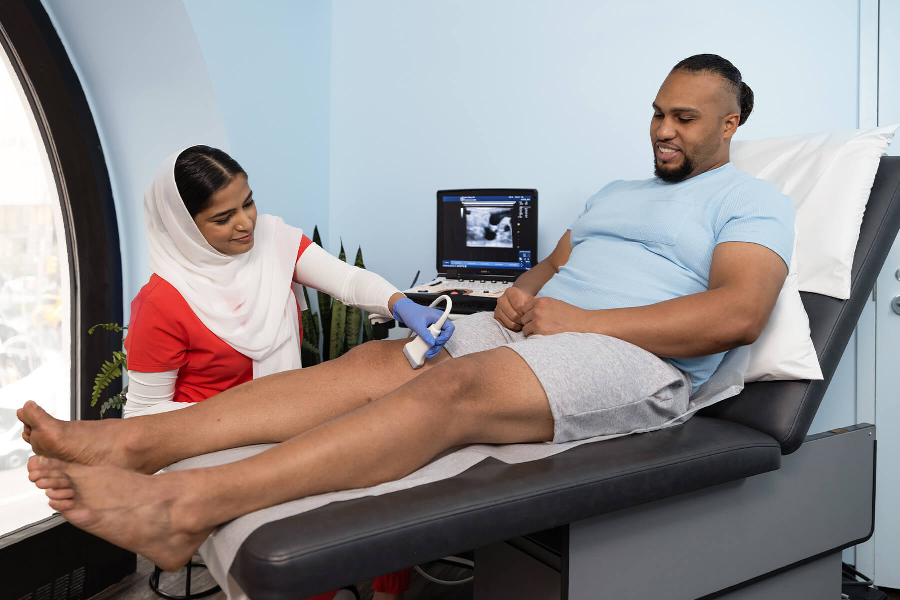
An ultrasound appointment for leg veins is completely painless and generally takes about 30 to 45 minutes total. Here is what you can expect from start to finish:
- Arrival & Positioning: You will check in, change into comfortable shorts or a gown, and lie down on an examination table. The exam is usually performed while you stand or tilt slightly to maximize blood pooling in the legs.
- Gel & Transducer Movement: The technician applies a warm, water-based ultrasound gel to your leg and glides a handheld probe (transducer) along the skin.
- Compression Checks: At various intervals, your vein specialist may gently squeeze your calf or thigh (or ask you to perform a breathing maneuver) to test how your vein valves respond to pressure changes.
- The Doppler Sound: You may hear a whooshing sound during the scan; this is normal and means that the Doppler technology is doing its job.
How to Prepare for Your Duplex Ultrasound
Little to no prepping is needed for a venous duplex scan, but a few quick steps will make your appointment even smoother:
- Wear loose, comfortable clothing that you can easily adjust or roll up above the knee.
- Skip lotions, oils, or creams on your legs the day of the scan, as they can interfere with the acoustic gel and sensor contact.
- Bring your prior imaging files or referral paperwork if you are visiting a new specialist.

Duplex Ultrasound Myths vs. Facts
Let’s clear up some common myths and misconceptions about venous ultrasound scans.
Myth: “Ultrasound uses harmful radiation.”
Fact: It does not. Duplex ultrasound uses harmless high-frequency sound waves only, so there’s no radiation and no contrast dye.
Myth: “It’s a painful procedure.”
Fact: The test is entirely non-invasive and painless; you will only feel light pressure from the handheld probe or manual compression.
Myth: “You only need a scan if your veins are visibly bulging.”
Fact: Not true. Significant venous reflux can occur deep beneath the surface with zero visible varicose veins, meaning ultrasound catches functional issues a standard physical exam misses.
Myth: “It’s the exact same as an obstetric pregnancy ultrasound.”
Fact: Yes and no. While they share core technology, vein scans use specialized Doppler modes to measure blood velocity and valve closure that standard pregnancy scans don’t require.
Myth: “If the superficial scan is clear, you don’t have vein disease.”
Fact: Not necessarily. A superficial scan does not rule out deep or perforator system issues; a complete diagnostic protocol evaluates all three systems to ensure nothing is missed.
Understanding Your Results and Next Steps
Once your scan is complete, your vein specialist will review the results. Normal findings mean your vein walls are flexible, blood moves efficiently in one direction, and your valves close tightly with no abnormal reflux or clotting.
Abnormal findings (such as reflux exceeding 0.5 seconds or a sluggish flow) can point to chronic venous insufficiency. These results are compared to a clinical framework called the CEAP classification to grade your condition’s severity.
Results are typically ready the same day or within a few days depending on the clinic. If reflux is confirmed, your vein specialist will work with you to choose the right minimally invasive treatment.

Getting Answers with a Duplex Ultrasound
A duplex ultrasound is a fast, entirely painless, and first-line test used by vein specialists. With structural imaging and blood-flow information, it gives your vein care team a clear picture of your vascular health. If you are experiencing heavy, aching legs or visible varicose veins, scheduling a free vein health assessment at one of Metro Vein Centers’ locations near you is the first step toward healthier, more comfortable legs.
Frequently Asked Questions
Plan for about 30 to 45 minutes total (roughly 15 minutes per leg), depending on how many segments your specialist evaluates.
Yes, insurance policies will typically cover venous duplex scans when they are considered medically necessary. Purely cosmetic screenings generally do not qualify. Always consult your provider or vein clinic to verify your insurance coverage.
Yes. Because the procedure is completely non-invasive, there is zero recovery time, downtime, or post-exam restriction.
If the scan reveals venous insufficiency or valve reflux, your specialist will review the findings with you and map out a personalized treatment plan tailored to your symptoms.
References
Alhejily, M., & Shapiro, M. (2023). Duplex ultrasound. StatPearls Publishing. https://www.ncbi.nlm.nih.gov/books/NBK459266/
Eberhardt, R. T., & Raffetto, J. D. (2014). Chronic venous insufficiency. Circulation, 130(4), 333–346. https://doi.org/10.1161/CIRCULATIONAHA.113.006898
Society for Vascular Surgery. (n.d.). Duplex ultrasound. https://vascular.org/your-vascular-health/your-care-journey/testing/duplex-ultrasound

Dr. Philip LoPresti
Meet Dr. Philip LoPresti, DABVLM, FACS, a board-certified surgeon and vein specialist and surgeon with over 20 years of experience. Schedule an appointment with him in Queens, NY today.
Meet Dr. Philip LoPresti
Trusted insight from the nationally accredited, board-certified vein doctors at Metro Vein Centers.